
Common facial fractures in southern California, tend to be orbital fractures, nasal bone fractures, and mandible fractures. The reconstructive treatment for many of these fractures requires the same care and planning as cosmetic procedures and with their own set of technical challenges.
With nasal bone fractures and the resulting deviated septum correction of the airway obstruction and cosmetic deformity is treated with rhinoplasty and airway surgery. Usually 6 months time should elapse until swelling subsides before surgical correction with rhinoplasty. In the case of orbital fractures treatment is usually much sooner.
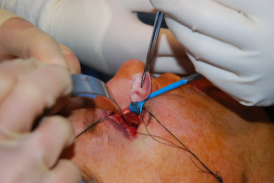
The orbit is comprised of seven bones: frontal, ethmoid, lacrimal, palatine, sphenoid, maxilla, zygoma. The floor and medial wall of the orbit the maxilla and ethmoid tend to be paper thin, having been termed, the lamina papyrecea. The design is truly brilliant, as force applied to the globe or eyeball is transmitted to this lamina papyrecia resulting in fracture of the lamina papyrecia preferentially before exceeding the pressure of the eye. There fore the body attempts to preserve vision at the expense of the bone. The floor may necessitate repair with titanium mesh, porous polypropylene, cranial bone graft, or cartilage. I have found that the contour of auricular or ear cartilage approximates very well the concavity of the medial wall or floor.
I prefer using the transconjuntival approach to prevent a visible scar on the face. If needed, I have learned that a lateral canthotomy provides excellent exposure to the floor, with this extension being readily hidden in one of the "crow's feet" of the lateral aspect of the face.