Thursday, September 30, 2010

Mandible Fractures. Symphysis Fractures


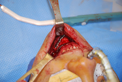
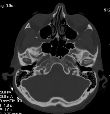
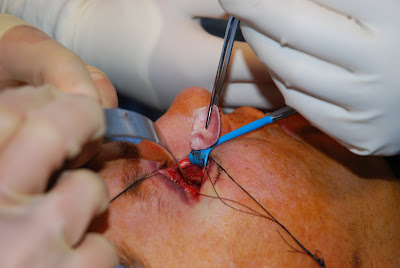
Mandible fractures are common and are some of the most enjoyable operations I perform. I prefer to treat many mandible fractures with open reduction and internal fixation, followed by a short course of intermaxillary fixation with Synthes, IMF screws.


Tuesday, September 28, 2010

Mandible Fractures


Mandible fractures are common. Typically, mandible fractures are best treated with open reduction and internal fixation (ORIF). ORIF involves using plates and screws to hold the fracture in position. This fixation allows arch bars or intermaxillary fixation to be removed sooner and allow the patient to use their jaw in a timelier manner.

www.drbriandickinson.com

Wednesday, July 7, 2010

Facial Fractures. Mandible Fractures


Mandible fractures or jaw fractures are a common occurrence. Fractures of the mandible are analogous to fractures of the pelvis in that fractures often occur in more than one location.

Depending on the textbooks that one reads, the frequency of occurrence of most mandible fractures is as follows: body (29%), condyle (26%), angle (25%), symphyses (17%), ramus (4%) and coronoid process (1%).

I prefer to treat most mandible fractures with open reduction and internal fixation with plates and screws as this affords the patient a more rapid return to jaw mobilization and can prevent stiffness at the temporomandibular joint.

Facial Fractures. Journal of Craniofacial Surgery


Pediatric Facial Fractures: Current National Incidence, Distribution, and Health Care Resource Use. Vyas, Raj M. MD; Dickinson, Brian P. MD; Wasson, Kristy L. BA; Roostaeian, Jason MD; Bradley, James P. MD
Journal of Craniofacial Surgery. 19(2):339-349, March 2008.

Wednesday, June 30, 2010

Zygoma Fractures, Orbital Fractures, Maxillary Fracture


Fractures of the zygomaticomaxillary complex frequently occur in concert with fractures of the orbital floor and orbital rim. Access to the these fractures is afforded thr0ugh exposure via the conjuntival incsion of the eye, the maxilary gingivobuccal sulcus, a lateral canthal extesion, and occasionally small access incisions on the cheek. Compliance post-operatively for the treatment of these fractures requires elevation of the head of bed to prevent edema, pain control, soft diet, and rest.

Facial Fractures. Nasal Bone Fractres


Nasal bone fractures are the most common types of facial fractures. Often in the emergency room the physician will examine the septum of the nose to make sure there is no septal hematoma. If the nasal bone fracture produces a deformity, then the plastic & reconstructive surgeon to performs closed or open reduction usually between 3 and 7 days, and up to 2 weeks after the injury. If possible and if the surgeon has the opportunity, appropriate reduction of the fracture can occur within the first several hours following the injury before significant edema has appeared. It is quite common following nasal trauma for patients to have airway obstruction and often necessitate airway surgery or corrective rhinoplasty approximately 6 months following their injury.

Tuesday, June 29, 2010

Facial Fractures. Zygomaticomaxillary Complex Fractures.


Zygomaticomaxillary fractures are common. Typically, these fractures occur when a person's head strikes the head, fist, or other solid object. The zygoma which protrudes a greater distance than the globe absorbs the impact at the expense of the eye.

Appropriate and optimal treatment of these fractures occurs within the first week of injury before the fracture has time to heal in incorrect alignment. Access if often gained to the inside of the orbit to ensure that the lateral orbital wall is correctly aligned.

http://www.drbriandickinson.com/

Tuesday, June 8, 2010

Orbital Fractures. Roxbury Clinic & Surgery Center


Orbital fractures are very common. There is an increasing frequency of orbital fractures being repaired at the Roxbury Clinic & Surgery Center in Beverly Hills, CA. Orbital fractures are becoming frequently more common in older individuals who may fall and suffer a trauma to the region of their eye.
By nature of the design of the globe and bony orbital frame, the bone tends to fracture first to prevent damage to the eye itself. Repair of the bony defect is often required for greater than 1 square cm defects and/or defects comprising greater than 50% of the floor of the orbit.
Patients who come to the Roxbury Clinic & Surgery Center in Beverly Hills to have their surgery are fortunate to have the expertise of Dr. Kami Parsa. Dr. Kami Parsa is a world renowned ophthalmologist with exceptional talent in oculoplastic and reconstructive surgery of the eye. Smaller orbital floor defects are often repaired with ear cartilage harvested from the ear, while larger defects or defects of the orbital rim often require repair with titanium mesh or porous polyethylene.

Nasal Bone Fractures.


Fractures of the nasal bones are very common. Typically these fractures can be "re-set" in the emergency room setting and then splinted. Frequently, patients who have nasal bone factures develop airway obstruction requiring airway surgery. The goals of airway surgery are to re-stablish patency of the airway through straigtening of the nasal septum, reduction of the size of the inferior turbiates, or stabilizing the internal, and external nasal valves.

Autologous cartilage from the septum, ear, or occasionally when these donor sites have already utilized or cannot provide cartilage of sufficient quality or quantity then rib graft is used instead.

http://www.drbriandickinson.com/

Facial Trauma: Orbital Fractures


Common facial fractures in southern California, tend to be orbital fractures, nasal bone fractures, and mandible fractures. The reconstructive treatment for many of these fractures requires the same care and planning as cosmetic procedures and with their own set of technical challenges.
With nasal bone fractures and the resulting deviated septum correction of the airway obstruction and cosmetic deformity is treated with rhinoplasty and airway surgery. Usually 6 months time should elapse until swelling subsides before surgical correction with rhinoplasty. In the case of orbital fractures treatment is usually much sooner.
The orbit is comprised of seven bones: frontal, ethmoid, lacrimal, palatine, sphenoid, maxilla, zygoma. The floor and medial wall of the orbit the maxilla and ethmoid tend to be paper thin, having been termed, the lamina papyrecea. The design is truly brilliant, as force applied to the globe or eyeball is transmitted to this lamina papyrecia resulting in fracture of the lamina papyrecia preferentially before exceeding the pressure of the eye. There fore the body attempts to preserve vision at the expense of the bone. The floor may necessitate repair with titanium mesh, porous polypropylene, cranial bone graft, or cartilage. I have found that the contour of auricular or ear cartilage approximates very well the concavity of the medial wall or floor.
I prefer using the transconjuntival approach to prevent a visible scar on the face. If needed, I have learned that a lateral canthotomy provides excellent exposure to the floor, with this extension being readily hidden in one of the "crow's feet" of the lateral aspect of the face.

Facial Fractures. Orbital Floor Fractures


Management of facial trauma and facial fractures are surgical procedures I find very rewarding. I find the anatomy fascinating and I enjoy continually trying to improve ways in which to conceal scars to manage these fractures.
Fractures of the orbital floor that comprise greater that 50% of the orbital floor, greater than 1 cm squared, or have evidence of clinical or radiographic entrapment of the extraocular muscles I often choose to repair with autologous ear cartilage or Synthes porous polyethylene.

Facial Fractures. Facial Trauma.


Surfing accidents may cause significant facial trauma. Over the past several months, the weather has been good, the water warm, and the frequency of surfing accidents has been high. The impact of a surfboard to the face can cause a lateral orbital wall and medial orbital wall blowout fracture as depicted here in the 3D CT-SCAN reconstruction. Synthes titanium microplates were uses to repair the lateral orbital wall fracture.
I have found that autologous ear cartilage is an excellent substitute to cranial bone graft in that the harvest is less traumatic and the curvature of the ear cartilage closely matches that of the medial orbital wall and orbital floor.

Friday, February 12, 2010

Orbital Fractures


Orbital fractures are very common. There is an increasing frequency of orbital fractures being repaired. Orbital fractures are becoming frequently more common in older individuals who may fall and suffer a trauma to the region of their eye.

By nature of the design of the globe and bony orbital frame, the bone tends to fracture first to prevent damage to the eye itself. Repair of the bony defect is often required for greater than 1 square cm defects and/or defects comprising greater than 50% of the floor of the orbit.

Smaller orbital floor defects are often repaired with ear cartilage harvested from the ear, while larger defects or defects of the orbital rim often require repair with titanium mesh or porous polyethylene.
Brian P. Dickinson, M.D.