Friday, January 25, 2013

SMAS Platsmal Coverage of Posterior Neck Wounds


Chronic wounds of the posterior neck can be covered and closed with large SMAS-platysmal flaps. Posterior neck wounds can occasionally occur from long-standing hardware or deep brain stimulators. Such hardware is important for patient function and is necessary to be maintained.

Mobilizing the skin, superficial musculoaponeurotic system (SMAS) and platysma posteriorly, can allow for well vascularized skin to cover wounds behind the ear.


www.drbriandickinson.com

Monday, September 24, 2012

"Blepharoplasty Graft"-Using Upper Lid Skin to Release Scar Contractures of the Face


Defects of the upper lip and nose can be challenging. Skin defects can result from trauma, surgery, etc. Loss of tissue in these areas can be challenging for both the physician and patient.
Scar that develops between structures that are mobile such as a joint (knee, elbow, finger, etc.) or on areas of the face can impair motion. As the scar contracts even more, motion can be further impaired and eventually further scarring can cause distortion of the normal position of these structures.
 
In order to allow mobility of these structures or restore them to their natural position, tissue must be borrowed or grafted from one area and placed in the area where the scar is excised. The skin of the upper lids can often be used to graft areas of the face as the color match and texture is often similar.


Sunday, June 24, 2012

Scalp Flaps to Close Forehead and Lateral Brow Wounds


Scalp flaps are commonly used to close defects on the scalp that result from tumors, trauma, radiation, etc.

Often these scalp flaps are used when tissue can be borrowed from one area and transferred to another, with the resulting donor site being easily skin grafted.

One of the more common scalp flaps that I use is the lateral scalp flap based on the superficial temporal artery to cover defects of the lateral aspect of the forehead or lateral brow region. It is important to draw the scalp flap extending more cephalad than would be expected as the flap tends to loose length as it is "arc'ed" anteriorly. Always remember to raise the flap in a supra-periosteal, or subgaleal plane at the location of the region to be skin grafted. This facilitates graft take.


www.drbriandickinson.com

Zygomaticomaxillary Complex Fractures

Zygomaticomaxillary complex fractures with significantly comminuted zygomatic arch fractures need wide exposure to the lateral aspect of the orbit as well as the zygomatic arch. This exposure is necessary to confirm adequate alignment of the lateral orbital wall as well as allow for stable open reduction and internal fixation.


I have found that the coronal exposure with subperiosteal elevation to the nasion with release of the supraorbital neurovascular bundles, bilaterally, can deliver excellent exposure of the lateral orbital rim. Once the operative surgeon has obtained this exposure, there is now easy access to plating the strongest fracture point of the zygomaticomaxillary complex. While the plate is being applied, pressure can be placed superiorly and laterally on the fractured complex, and fracture position can also be confirmed at the orbital rim.


Once the zygomaticofrontal process is stabilized, I then turn my attention to the zygomatic arch. Great care is taken when approaching the zygomatic arch, so that the frontal branch of the facial nerve can be elevated in the flap. Typically, I follow the zygomaticofrontal process inferiorly with my periosteal elevator, and, once the superficial temporal fat pad is reached, I push the fat pad inferiorly and laterally. This confirms not only that I am approaching the arch at a level amenable to plating, but also the frontal branch of the facial nerve is protected.


www.drbriandickinson.com

Thursday, September 30, 2010

Mandible Fractures. Symphysis Fractures


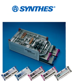
Mandible fractures are common and are some of the most enjoyable operations I perform. I prefer to treat many mandible fractures with open reduction and internal fixation, followed by a short course of intermaxillary fixation with Synthes, IMF screws.


Tuesday, September 28, 2010

Mandible Fractures


Mandible fractures are common. Typically, mandible fractures are best treated with open reduction and internal fixation (ORIF). ORIF involves using plates and screws to hold the fracture in position. This fixation allows arch bars or intermaxillary fixation to be removed sooner and allow the patient to use their jaw in a timelier manner.

www.drbriandickinson.com